


This is the picture and labeling of the sagital cut of the brain.


Cerebrum is yellow, brain stem is red, cerebellum is green.
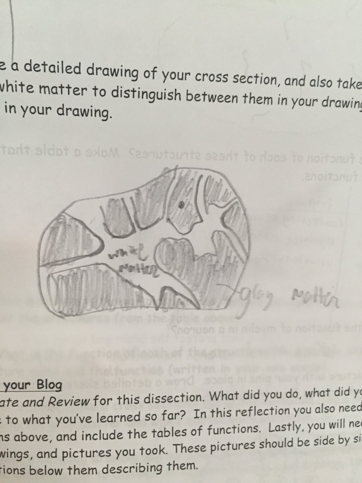
In this lab we labeled and made incisions on the sheep brain. There was only two cuts we had to make. one was a sagittal cut and than a cross section cut on the cerebrum. we labeled many parts of the brain starting with discovering the anterior and posterior side of it. After that we got the cerebrum and the cerebellum which is the bottom part that is shaped like a bell. The brain stem was really fragile. Once we made the sagital cut labeling got a little more difficult, we labeled the thalamus, optic nerve, medulla oblongata, pons, mid brain, corpus callosum and hypothalamus. Than we had to make a cross section on the cerebrum to make the white and gray matter more distinct.
Brain stem
|
The Brain stem connects neurons to the brain and is assosiated with many involuntary movements in the brain.
|
Cerebellum
|
coordinates voluntary movements and receieves sensory info from the sensory receptors.
|
Cerebrum
|
This is related to the thughts and the action of the body, the voluntary movements.
|
Thalamus
|
Relay sensory and motar signals, regulate consisness alertness and sleep.
|
Optic nerve
|
transmits information from the retina of the eye to the brain
|
Medulla oblongata
|
Controls body functions, coordination of body movements.
|
pons
|
Relay info from the cerebrum to the cerebellum
|
midbrain
|
midbrain
|
Corpus callosum
|
Helps share information
|
hypothalamus
|
Maintain homeostasis
|
The function of the myelin is to wrap around axons and helps the impulse flow through.
No comments:
Post a Comment